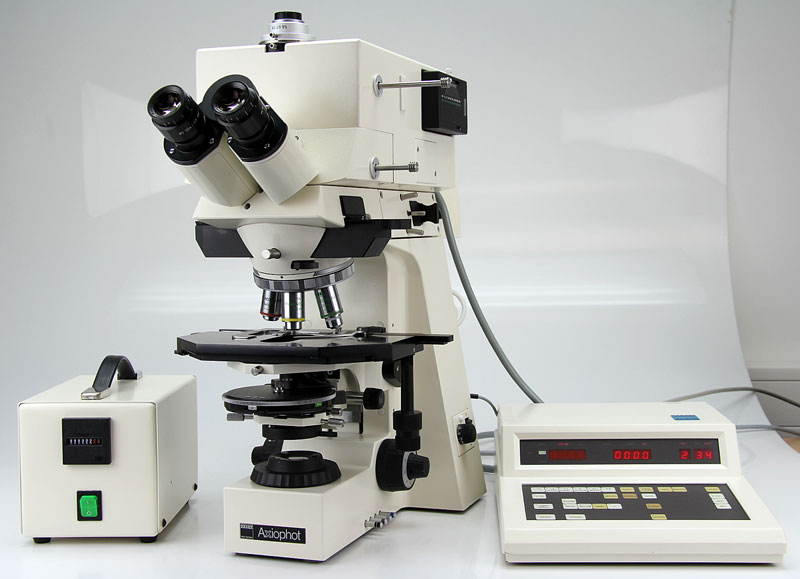
Zeiss Axiophot
Pricing
| $36/Hour | TAMU Users & Federal Government Agency Users |
| $31/Hour | Off-Peak & Automated Runs (TAMU Users & Federal Government Users only) 5:00 p.m. - 8:00 a.m. |
| $90/Hour | Other Universities |
| $135/Hour | Industry |
Capabilities:
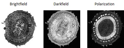
This light microscope is equipped for bright field (transmitted and reflected), phase contrast, transmitted and reflected polarization, Nomarski differential interference contrast microscopy.
Specifications:
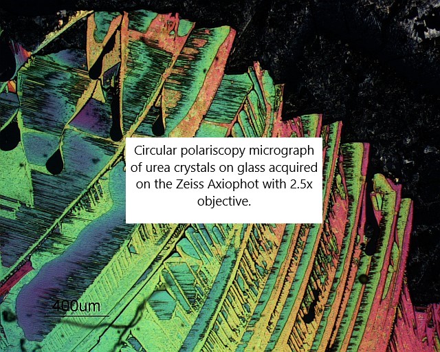
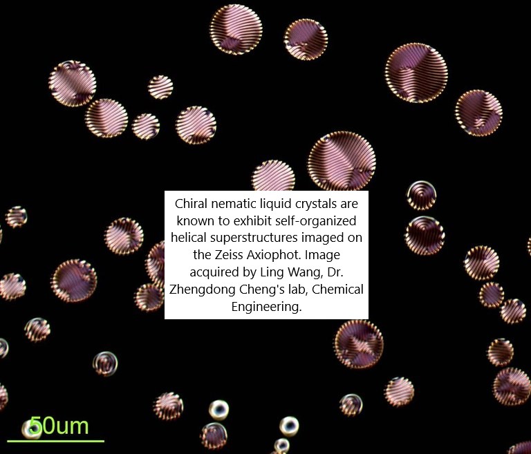
Imaging Modes: DIC (reflected and transmitted), phase contrast, brightfield, darkfield, polarization (reflected and transmitted)
Objectives:
- 5X, 10X, 20X, and 40X dry objectives
- 63X/1.40 and 100X/1.3 oil immersion objectives
Camera: Color CMOS camera
Stage: Motorized focus via Prior Optiscan system which allows acquisition of stacks
Software: Image acquisition is controlled by the free Micro-Manager software
For imaging services, training, and questions
Please contact Dr. Stanislav Vitha, stanvitha@tamu.edu
Helpful Hints:
- Download Training Policy: Axiophot Training Policy
- Download User Guide: Zeiss-Axiophot-manual.pdf
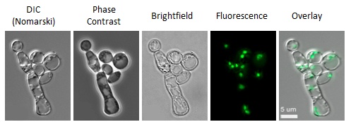
Examples of imaging modes:
Example 1:
Live cells of the filamentous fungus Neurospora crassa, expressing Green Fluorescent Protein (GFP) fused to Histone H1. The GFP fusion protein is localized in the nucleus.
Example 2:
Cross-section of a wooded plant stem.